
Page 809 - TNFlipTest
P. 809
Toronto Notes 2019
CNS Tumours
Neurosurgery NS11
CNS Tumours
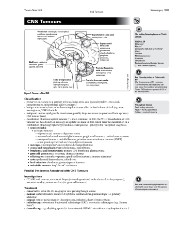
Ventricular: colloid cyst, choroid plexus papilloma, ependymoma,
germinoma, teratoma,
meningioma
Supratentorial extra-axial:
meningioma, cysts
Supratentorial intra-axial: astrocytoma, glioblastoma, oligodendroglioma, ganglioma, lymphoma, metastases
Posterior fossa intra- axial: schwannoma, meningioma, cysts, metastases
Posterior fossa extra-axial:
schwannoma, meningioma, cyst, metastases
DDx for Ring Enhancing Lesion on CT with Contrast
MAGICAL DR
Metastases*
Abscess*
Glioblastoma (high grade astrocytoma)* Infarct
Contusion
AIDS (toxoplasmosis)
Lymphoma
Demyelination
Resolving hematoma, Radiation Necrosis (*3 most common diagnoses)
Ring Enhancing Lesions in Patients with HIV
DDx: Toxoplasmosis or CNS Lymphoma
Tx: Pyrimethamine and Sulfadiazine; later brain biopsy if no resolution with antimicrobial Primary CNS lymphoma reported in 6-20% of HIV infected patients
Primary Brain Tumours
Rarelyundergometastasis Adults = mostly supratentorial Children = mostly infratentorial
Skull base: carcinoma, chordoma, glomus jugulare, osteoma
Figure 9. Tumours of the CNS
Classification
Sellar or suprasellar:
pituitary adenoma, craniopharyngioma, optic nerve glioma, cyst
• primaryvs.metastatic(e.g.primaryinbreast,lung),intra-axial(parenchymal)vs.extra-axial, supratentorial vs. infratentorial, adult vs. pediatric
• benign:non-invasive,butcanbedevastatingduetomasseffectinfixedvolumeofskull(e.g.most meningiomas, WHO Grade I)
• malignant:impliesrapidgrowth,invasiveness,possiblydrop-metastasestospinalcordfromaprimary CNS tumour (rare)
• classificationofnervoussystemtumours(*=mostcommon).In2007,theWHOClassificationofCNS tumours was based solely on histology; an update was made in 2016 which bases the classification on a combination of histology (phenotype) and molecular genetics (genotype) for “integrated” diagnoses:
■ neuroepithelial
◆ astrocytic tumours
– oligoastrocytic tumours: oligoastrocytoma
– neuronal and mixed neuronal-glial tumours: ganglion cell tumours, cerebral neurocytomas – embryonal tumours: medulloblastoma, primitive neuroectodermal tumours (PNET)
– other: pineal, ependymal, and choroid plexus tumours
■ meningeal: meningiomas*, mesenchymal, hemangioblastomas
■ cranialandparaspinalnerves:schwannoma,neurofibroma
■ lymphomasandhematopoietic:primaryCNSlymphoma,plasmacytoma
■ germ cell: germinomas, teratomas, choriocarcinomas
■ sellar region: craniopharyngiomas, spindle cell oncocytoma, pituitary adenomas* ■ cysts: epidermoid/dermoid cysts, colloid cysts
■ local extension: chordomas, glomus jugulare tumours
■ metastatic tumours: lung*, breast*, melanoma
Familial Syndromes Associated with CNS Tumours
Investigations
• CT,MRIwithcontrast,stereotacticbiopsy(tissuediagnosisandmolecularmarkersforprognosis), metastatic workup, tumour markers (i.e. germ cell tumours)
Treatment
• conservative:serialHx,Px,imagingforslowgrowing/benignlesions
• medical:corticosteroidstoreduceICP,cytotoxiccerebraledema,pharmacologic(i.e.pituitary
adenoma)
• surgical:totalorpartialexcision(decompressive,palliative),shuntifhydrocephalus
• radiotherapy:conventionalfractionatedradiotherapy(XRT),stereotacticradiosurgery(e.g.Gamma
Knife®)
• chemotherapy:e.g.alkylatingagents(i.e.temozolomide(GBM)*,vincristine,cyclophosphamide,etc.)
New onset communicating hydrocephalus in a patient with cancer should raise the suspicion of leptomeningeal carcinomatosis
© Ryan Kissinger 2010
