
Page 807 - TNFlipTest
P. 807
Toronto Notes 2019 Hydrocephalus
• morbidity:riskofblindness,whichisnotreliablycorrelatedtoduration,symptomsorclinicalcourse
• clinicalcourse:usuallyself-limited,recurrencein10%,chronicinsome
Investigations
• MRI-brain(withandwithoutcontrast):slitlikeventriclesanddistendedperiopticsubarachnoidspace, but otherwise normal
■ rule out: venous sinus thrombosis, mass, infection, hydrocephalus
• LPfindings
■ openingpressure>25cmH2O
■ normal CSF analysis
• ophthalmologic:fields,acuity,papilledema
Treatment
• lifestylechange:encourageweightloss,fluid/saltrestriction
• pharmacotherapy:acetazolamide(decreasesCSFproduction),thiazidediuretic,orfurosemide;
discontinue offending medications
• surgery:ifabovefail,serialLPs(temporizing),shunts,opticnervesheathfenestration(ifprogressive
impairment of visual acuity)
• longterm:2yrfollow-up,repeatimagingtoruleoutocculttumour,ophthalmologyfollow-up
Hydrocephalus
• forhydrocephalusinchildren,seePediatricNeurosurgery,NS35
Definition
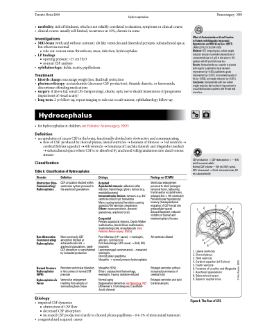
• accumulationofexcessCSFinthebrain,functionallydividedintoobstructiveandcommunicating
■ flow of CSF: produced by choroid plexus, lateral ventricles → foramen of Monroe → 3rd ventricle →
cerebral/Sylvian aqueduct → 4th ventricle → foramina of Luschka (lateral) and Magendie (medial) → subarachnoid space where CSF is re-absorbed by arachnoid villi/granulations into dural venous sinuses
Neurosurgery NS9
Effect of Acetazolamide on Visual Function in Patients with Idiopathic Intracranial Hypertension and Mild Visual Loss (IIHTT) JAMA 2014;311(16):1641-1651
Methods: RCT comparing low-sodium weight- reduction diet plus maximally tolerated dose of acetazolamide (up to 4 g/d) to diet alone in 165 patients with IIH and mild visual loss.
Results: Acetazolamide was superior to placebo with regards to perimetric mean deviation improvement (p=0.05), papilledema grade improvement (p<0.001), vision-related quality of life (p=0.003), and weight reduction (p<0.001). Conclusion: Acetazolamide with low-sodium weight-reduction diet resulted in improvement in visual field function in patients with IIH and mild visual loss.
Classification
Table 6. Classification of Hydrocephalus
CSF production = CSF reabsorption = ~ 500 mL/d in normal adults
Normal CSF volume ~150 mL (50% spinal, 50% intracranial → 25 mL intraventricular, 50 mL subarachnoid)
Disorder
Obstructive (Non- Communicating) Hydrocephalus
Non-Obstructive (Communicating) Hydrocephalus
Normal Pressure Hydrocephalus (NPH)
Hydrocephalus Ex Vacuo
Etiology
Definition
CSF circulation blocked within ventricular system proximal to the arachnoid granulations
Most commonly CSF absorption blocked at extraventricular site = arachnoid granulations, rarely CSF absorption is overwhelmed by increased production
Persistent ventricular dilatation in the context of normal CSF pressure
Ventricular enlargement resulting from atrophy of surrounding brain tissue
Etiology
Acquired
Aqueductal stenosis: adhesions after infection, hemorrhage; gliosis, tumour (e.g. medulloblastoma)
Intraventricular lesions: tumours, e.g. 3rd ventricle colloid cyst, hematoma
Mass causing tentorial herniation causing aqueduct/4th ventricle compression Others: neurosarcoidosis, abscess/ granulomas, arachnoid cysts
Congenital
Primary aqueductal stenosis, Dandy-Walker malformation, Arnold-Chiari malformation, myelomeningocele, encephalocele (see Pediatric Neurosurgery, NS35)
Post-infectious (#1 cause) → meningitis, abscess, cysticercosis
Post-hemorrhagic (#2 cause) → SAH, IVH, traumatic
Leptomeningeal carcinomatosis – metastatic meningitis
Choroid plexus papilloma
Idiopathic → normal pressure hydrocephalus
Idiopathic (50%)
Others: subarachnoid hemorrhage, meningitis, trauma, radiation-induced
Normal aging
Degenerative dementias see Neurology, N21 (Alzheimer’s, frontotemporal, Creutzfeldt- Jacob disease)
Findings on CT/MRI
Ventricular enlargement
proximal to block (enlarged
temporal horns, ballooning
frontal and/or occipital horns,
enlarged 3rd ± 4th ventricles)
Periventricular hypodensity/
lucency (transependymal
migration of CSF forced into 2 3 extracellular space) 1
7
9
Sulcal effacement, reduced visibility of Sylvian and interhemispheric fissures
All ventricles dilated
Enlarged ventricles without increased prominence of cerebral sulci
Enlarged ventricles and sulci Cerebral atrophy
8
4
56
• impairedCSFdynamics
■ obstruction of CSF flow
■ decreased CSF absorption
■ increased CSF production (rarely in choroid plexus papilloma – 0.4-1% of intracranial tumours)
• congenitalandacquiredcauses
1. Lateral ventricles
2. Choroid plexus
3. Third ventricle
4. Cerebral aqueduct (of Sylvius) 5. Fourth ventricle
6. Foramina of Luschka and Magendie 7. Arachnoid granulations
8. Subarachnoid space
9. Superior sagittal sinus
Figure 8. The flow of CFS
© Kari Francis 2004
