
Page 808 - TNFlipTest
P. 808
NS10 Neurosurgery
Classic (Hakim’s) Triad of NPH Progression
“Wet, wacky, wobbly”: Incontinence, dementia, ataxia
Important Features to Note on CT and MRI (± contrast enhancement)
• Lesions (± edema, necrosis, hemorrhage)
• Midline shifts and herniations
• Effacement of ventricles and sulci (often ipsilateral), basal cisterns
• Single or multiple (multiple implies metastasis)
Hydrocephalus Toronto Notes 2019 • estimatedprevalence1-1.5%;incidenceofcongenitalhydrocephalus~1-2/1,000livebirths
Clinical Features
• acutehydrocephalus:signsandsymptomsofacutelyelevatedICP(seeTable3)
• chronic/gradualonsethydrocephalus:(wktomo;i.e.NPH)presentswithaclassictriad(Hakim’sTriad)
■ Ataxia(magneticgait)+apraxia(pressureofventricleonlowerextremitymotorfibres→gait disturbance)
■ Incontinence(pressureoncorticalbowel/bladdercentre)
■ Dementia(subcortical)
Investigations
• imaging
• CT/MRIfindings(seeTable6)
• ultrasound(throughanteriorfontanelleininfants):ventriculomegaly,sizeandlocationoflesions(e.g.
IVH)
• mantleradionuclide cisternography can test CSF flow and absorption rate (unreliable)
• ICPmonitoring(e.g.LP,EVD)maybeusedtoinvestigateNPHandtestresponsetoshunting(lumbar
tap test)
Treatment
• externalventriculardrain(EVD)
• intermittentLPsfortransientcommunicatinghydrocephalus(SAH,IVHinprematureinfants)
• eliminatingobstruction(i.e.excisionofmass,posteriorfossadecompressionforChiariMalformation) • endoscopic
■ endoscopic third ventriculostomy (ETV) ± choroid plexus cauterization (for obstructive hydrocephalus)
■ endoscopic placement of aqueductal stent • shunt
■ ventriculoperitoneal (VP): most common shunt
■ ventriculopleural (VPl)
■ ventriculoatrial (VA)
■ lumboperitoneal: for communicating hydrocephalus and pseudotumour cerebri
Epidemiology
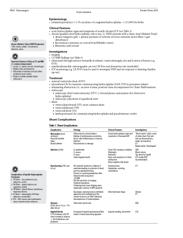
Shunt Complications
Table 7. Shunt Complications
Complications of Specific Hydrocephalus Treatments
1. VP Shunt – intra-abdominal cysts,
adhesions, ascites
2. VA Shunt – greater infection risk,
septicemia, emboli
3. VPl Shunt – pleural effusion, hydrothorax,
respiratory distress
4. LP Shunt – radiculopathy, CSF leaks,
adhesions, arachnoiditis
5. ETV – 56% success rate, hypothalamic
injury, traumatic basilar aneurysm
Complication
Obstruction (most common) Proximal Catheter Valve
Distal Catheter Infection (3-6%)
Overshunting (10% over 6.5 yr)
Seizures
(5.5% risk in 1st yr, 1.1% after 3rd yr)
Inguinal Hernia
(17% incidence with VP shunt inserted in infancy) ± skin breakdown over hardware
Etiology
Obstruction by choroid plexus Buildup of proteinaceous accretions, blood, cells (inflammatory or tumour) Infection
Disconnection or damage
S. epidermidis
S. aureus
P. acnes Gram-negative bacilli
Slit ventricle syndrome, collapse of ventricles leading to occlusion of shunt ports by ependymal lining
Chronic or recurring headaches often relieved when lying down
CT/MRI
Slit-like ventricles on imaging Subdural hematoma
Collapsing brain tears bridging veins (especially common in NPH patients)
Secondary craniosynostosis (children): apposition and overlapping of the cranial sutures in an infant following decompression of hydrocephalus
Ventricular shunts only
Increased intraperitoneal pressure/fluid results in hernia becoming apparent
Clinical Features
Acute hydrocephalus signs and symptoms of Increased ICP
Fever, N/V, anorexia, irritability Meningitis
Peritonitis
Signs and symptoms of shunt obstruction
Shunt nephritis (VA shunt)
Asymptomatic Headaches, vomiting, somnolence
Abnormal head shape
Inguinal swelling, discomfort
Investigations
“Shunt series” (plain x-rays of entire shunt that only rule-out disconnection, break, tip migration)
CT
Radionuclide “shuntogram”
CBC
Blood culture
Tap shunt for CandS (LP usually NOT recommended)
CT
Clinical CT
EEG U/S
