
Page 805 - TNFlipTest
P. 805
Toronto Notes 2019
Herniation Syndromes
Neurosurgery NS7
Clinical Features
Table 3. Clinical Features of Elevated ICP
Blood Brain Barrier
Glucose and amino acids cross slowly Non-polar/lipids cross fast
Infarction/neoplasm → destroy tight junctions → vasogenic edema
Cushing’s Triad of Acute Raised ICP
(full triad seen in 1/3 of cases) • Hypertension
• Bradycardia (late finding) • Irregular respiratory pattern
Papilledema
• Optic disc swelling with blurred margins (most commonly bilateral)
Clinical Features
Headache
Nausea and Vomiting
Level of Consciousness
GCS
Optic Disc Changes Visual Changes
Extra-Occular Movements
Herniation Syndromes Neurologic Deficits
Investigations
Acutely Elevated ICP
Chronic Progressive ICP Elevation
Both aggravated by stooping, coughing, and straining
Morning headaches: vasodilatation due to increased CO2 with recumbency
Present in both, though greater predilection in acutely elevated ICP
Lethargy if ICP = dBP or midbrain compression
Significant decline in GCS
Best index to monitor progress and predict outcome of acute intracranial process (see Neurotrauma, NS30)
Subtle changes suggesting papilledema (subtle elevations in disc margin, mild disc hyperemia) ± retinal hemorrhages (may take 24-48 h to develop)
Less common. Often not affected initially, however visual obscurations, flickering, or blurring can occur
Less common. CN VI palsy: due to long intracranial course, more sensitive to ICP changes and thus earlier sign of acutely increased ICP
Often falsely localizing (causative lesion remote to nerve) Upward gaze palsy and sunset eyes (especially in children with obstructive hydrocephalus)
Often occur
Focal deficits present
Irritability, inattentiveness. Normal or modestly reduced LOC, confusion
Can be unchanged or modestly decreased Obvious papilledema
Optic atrophy/blindness due to chronic papilledema
Enlarged blind spot, if advanced → episodic constrictions of visual fields (“grey-outs” lasting ~20 min)
Differentiate from papillitis (usually unilateral with decreased visual acuity)
Often full extraocular movement
Present if acute on chronic presentation Focal deficits can be present
• Larger blind spot
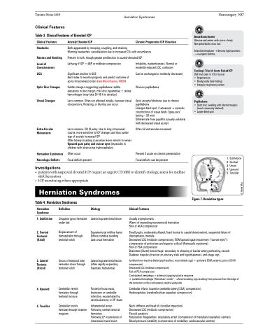
1
3 2 45
1. Subfalcine 2. Central
3. Uncal
4. Upward 5. Tonsillar
• patientswithsuspectedelevatedICPrequireanurgentCT/MRItoidentifyetiology,assessformidline shift/herniation
• ICPmonitoringwhereappropriate
Herniation Syndromes
Table 4. Herniation Syndromes
Figure 7. Herniation types
Herniation Syndrome
1. Subfalcine
2. Central Tentorial (Axial)
3. Lateral Tentoria (Uncal)
4. Upward
5. Tonsillar
Definition
Cingulate gyrus herniates under falx
Displacement of diencephalon through tentorial notch
Uncus of temporal lobe herniates down through tentorial notch
Cerebellar vermis herniates through tentorial incisura
Cerebellar tonsils herniate through foramen magnum
Etiology
Lateral supratentorial lesion
Supratentorial midline lesion Diffuse cerebral swelling Late uncal herniation
Lateral supratentorial lesion (often rapidly expanding traumatic hematoma)
Posterior fossa mass, brainstem or cerebellar infarction, exacerbated by ventriculostomy or VP shunt
Infratentorial lesion Following central tentorial herniation
Following LP in presence of intracranial mass lesion
Clinical Features
Usually asymptomatic
Warns of impending transtentorial herniation Risk of ACA compression
Small pupils, moderately dilated, fixed (rostral to caudal deterioration), sequential failure of diencephalon, medulla
Decreased LOC (midbrain compression), EOM/upward gaze impairment (“sunset eyes”): compression of pretectum and superior colliculi (Parinaud’s syndrome)
Risk of PCA compression
Brainstem (Duret) hemorrhage: secondary to shearing of basilar artery perforating vessels Diabetes insipidus (traction on pituitary stalk and hypothalamus), end-stage sign
Ipsilateral non-reactive dilated pupil (earliest, most reliable sign) + ipsilateral EOM paralysis, ptosis (CN III compression)
Decreased LOC (midbrain compression)
Risk of PCA compression
Contralateral hemiplegia ± extensor (upgoing) plantar response
± ipsilateral hemiplegia (“Kernohan’s notch” – a false localizing sign resulting from pressure from the edge of the tentorium on the contralateral cerebral peduncle)
Cerebellar infarct (superior cerebellar artery [SCA] compression) Hydrocephalus (cerebral/sylvian aqueduct compression)
Neck stiffness and head tilt (tonsillar impaction)
Decreased LOC (midbrain compression)
Flaccid paralysis
Respiratory irregularities, respiratory arrest (compression of medullary respiratory centres) Blood pressure instability (compression of medullary cardiovascular centres)
© Heidi Maj
