
Page 803 - TNFlipTest
P. 803
Toronto Notes 2019 Intracranial Pressure Dynamics Table 2. Consequences of Common Brain Lesions
Neurosurgery NS5
Location of Lesion
Frontal Lobe
Usually large lesions produce symptoms
Frontal Eye Fields
Broca’s Area
Posterior inferior frontal gyrus of dominant hemisphere
Occipital Lobe
Parietal Lobe
Either side
Dominant side (Left) Non-dominant side (Right)
Temporal Lobe
Wernicke’s Area
Posterior superior temporal gyrus of dominant hemisphere
Basal Ganglia
Subthalamic Nucleus Brainstem
Cerebellar Hemisphere Cerebellar Vermis
Consequence
Abulia, disinhibition, apathy, executive dysfunction, deficits in orientation and judgment, ± primitive reflex re-emergence, ± contralateral upper motor neuron signs (upgoing Babinski reflex and pronator drift)
Gaze deviation toward side of a destructive lesion Gaze deviation away from irritative lesion (i.e. seizure)
Non-fluent, dysarthric, aphasia Repetition impaired Comprehension spared
Contralateral homonymous hemianopsia
Dressing apraxia, cortical sensory loss, lower homonymous quadrantanopia Inattention or extinction of non-dominant side
Aphasias, Gerstmann’s syndrome
Hemispatial neglect, apraxias, agnosias (if temporal involvement)
Hippocampus: anterograde amnesia Upper homonymous hemianopia Wernicke’s aphasia (if left/dominant side)
Fluent aphasia Repetition impaired Comprehension impaired
Resting tremor
Chorea
Athetosis
Hemiplegia if internal capsule involved
Contralateral hemiballismus
Absent brainstem reflexes: oculocephalic, oculovestibular, corneal, gag, and cough Dorsal midbrain/pineal gland: Parinaud’s syndrome (supranuclear upward gaze palsy)
Pons: locked-in syndrome
Below red nucleus: decerebrate posture
Above red nucleus: decorticate posture
Reticular activating system (midbrain): reduced level of arousal
Cerebellar pontine angle: disequilibrium, ataxia, and other CN V,VII,VIII deficits
Intention tremor Ipsilateral limb ataxia
Fall towards side of lesion
Truncal ataxia Dysarthria
ICP/Volume Relationship
• Monro-KellieDoctrine:thebrainisencasedinarigidskullwithconstantintracranialvolume consisting of CSF, blood, and brain
• theincreaseinoneconstituentwill:1)necessitatetheredistributionofCSF,blood,and/orbrainand 2) increase ICP
• compensatorymechanismsinitiallymaintainanormalICP
• compensatoryreserve(spatialcompensation):60-80mLinyoungpeople,100-140mLinelderly
(largely due to cerebral atrophy)
■ immediate:egressionofCSFthroughforamenmagnumtospinalcanal,displacementofvenous
blood from sinuses into jugular veins
• oncecompensationisexhausted,ICPrisesexponentially:
■ late: displacement of arterial blood (decreased CPP) eventually leading to ischemia, increasing brain edema, or expanding mass displaces parenchyma into compartments under less pressure (Table 3)
■ end:cessationofcerebralperfusionwhenICP>MAP,cerebralherniationdownintoforamen magnum
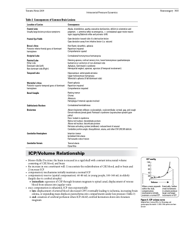
ICP mmHg
100
80
60
40
20
0
Volume
When a mass expands within the skull, compensatory mechanisms initially maintain a normal ICP.
Eventually, further small increments in volume produce larger and larger increments in ICP.
Figure 5. ICP volume curve
Adapted from: Lindsay KW, et al. Neurology and neurosurgery illustrated. © 2004. With permission from Elsevier
