
Page 815 - TNFlipTest
P. 815
Toronto Notes 2019 Blood Extradural (“Epidural”) Hematoma
Etiology
Neurosurgery NS17
• temporal-parietalskullfracture:85%areduetorupturedmiddlemeningealartery;remainderofcases are due to bleeding from middle meningeal vein, dural sinus, or bone/diploic veins
Epidemiology
• youngadult,M>F=4:1;rarebeforeage2orafterage60 • 1-4%oftraumaticheadinjuries
Clinical Features
• classicsequence(seenin<30%):post-traumaticreducedLOC,alucidintervalofseveralhours,then obtundation, hemiparesis, ipsilateral pupillary dilatation, and coma
• signsandsymptomsdependonseveritybutcanincludeH/A,N/V,amnesia,alteredLOC,aphasia, seizures, HTN, and respiratory distress
• deteriorationcantakehourstodays
Investigations
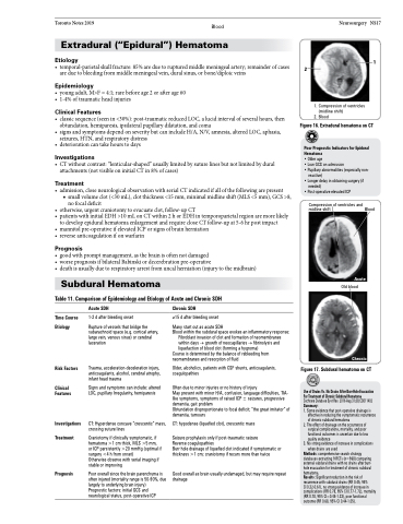
• CTwithoutcontrast:“lenticular-shaped”usuallylimitedbysuturelinesbutnotlimitedbydural attachments (not visible on initial CT in 8% of cases)
Treatment
• admission,closeneurologicalobservationwithserialCTindicatedifallofthefollowingarepresent
■ small volume clot (<30 mL), clot thickness <15 mm, minimal midline shift (MLS <5 mm), GCS >8,
no focal deficit
• otherwise, urgent craniotomy to evacuate clot, follow-up CT
• patientswithinitialEDH>10mLonCTwithin2horEDHintemporoparietalregionaremorelikely
to develop epidural hematoma enlargement and require close CT follow-up at 5-6 hr post impact
• mannitolpre-operativeifelevatedICPorsignsofbrainherniation
• reverseanticoagulationifonwarfarin
Prognosis
• goodwithpromptmanagement,asthebrainisoftennotdamaged
• worseprognosisifbilateralBabinskiordecerebrationpre-operative
• deathisusuallyduetorespiratoryarrestfromuncalherniation(injurytothemidbrain)
Subdural Hematoma
Table 11. Comparison of Epidemiology and Etiology of Acute and Chronic SDH
1
2
2. Blood
1. Compression of ventricles (midline shift)
Figure 16. Extradural hematoma on CT
Poor Prognostic Indicators for Epidural Hematoma
• Older age
• Low GCS on admission
• Pupillary abnormalities (especially non- reactive)
• Longer delay in obtaining surgery (if needed)
• Post-operative elevated ICP
Compression of ventricles and midline shift
Blood
Time Course Etiology
Risk Factors
Clinical Features
Investigations Treatment
Prognosis
Acute SDH
1-2 d after bleeding onset
Rupture of vessels that bridge the subarachnoid space (e.g. cortical artery, large vein, venous sinus) or cerebral laceration
Trauma, acceleration-deceleration injury, anticoagulants, alcohol, cerebral atrophy, infant head trauma
Signs and symptoms can include: altered LOC, pupillary Irregularity, hemiparesis
CT: Hyperdense concave “crescentic” mass, crossing suture lines
Craniotomy if clinically symptomatic, if hematoma >1 cm thick, MLS >5 mm, or ICP persistently >20 mmHg (optimal if surgery <4 h from onset)
Otherwise observe with serial imaging if stable or improving
Poor overall since the brain parenchyma is often injured (mortality range is 50-90%, due largely to underlying brain injury)
Prognostic factors: initial GCS and neurological status, post-operative ICP
Chronic SDH
≥15 d after bleeding onset Many start out as acute SDH
Blood within the subdural space evokes an inflammatory response: Fibroblast invasion of clot and formation of neomembranes within days → growth of neocapillaries → fibrinolysis and liquefaction of blood clot (forming a hygroma)
Course is determined by the balance of rebleeding from neomembranes and resorption of fluid
Older, alcoholics, patients with CSF shunts, anticoagulants, coagulopathies
Often due to minor injuries or no history of injury
May present with minor H/A, confusion, language difficulties, TIA- like symptoms, symptoms of raised ICP ± seizures, progressive dementia, gait problem
Obtundation disproportionate to focal deficit; “the great imitator” of dementia, tumours
CT: hypodense (liquefied clot), crescentic mass
Seizure prophylaxis only if post-traumatic seizure
Reverse coagulopathies
Burr hole drainage of liquefied clot indicated if symptomatic or thickness >1 cm; craniotomy if recurs more than twice
Good overall as brain usually undamaged, but may require repeat drainage
Acute
Old blood
Chronic
Figure 17. Subdural hematoma on CT
Use of Drains Vs. No Drains After Burr-Hole Evacuation For Treatment of Chronic Subdural Hematoma Cochrane Database Syst Rev. 2016 Aug 31;(8):CD011402 Summary:
1. Some evidence that post-operative drainage is effective in reducing the symptomatic recurrence of chronic subdural hematoma
2. The effect of drainage on the occurrence of surgical complications, mortality, and poor functional outcomes is uncertain due to low quality evidence
3. No strong evidence of increase in complications when drains are used
Methods: comprehensive search strategy databases extracting 9 RCTs (n=968) comparing external subdural drains with no drains after burr- hole evacuation for treatment of chronic subdural hematoma.
Results: Significant reduction in the risk of recurrence with subdural drains (RR 0.45, 95%
CI 0.32-0.61), no strong evidence of increase in complications (RR 0.78, 95% CI 0.77-1.72), mortality (RR 0.78, 95% CI=0.45-1.33), poor functional outcome (RR 0.68, 95% CI 0.44-1.05).
