
Page 814 - TNFlipTest
P. 814
NS16 Neurosurgery
Blood Toronto Notes 2019
3
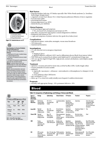
Figure 15. Cerebral abscess on CT
Recommendations For Duration Of Antibiotic Therapy For Brain Abscesses
Int J Infect Dis 2010 Oct;14 Suppl 4:S79-92 Summary:
1
2
Risk Factors
• lungabnormalities(infection,AVfistulas;especiallyOsler-Weber-Rendusyndrome[i.e.hereditary hemorrhagic telengiectasia])
• congenitalcoronaryheartdisease:R-to-Lshuntbypassespulmonaryfiltrationofmicro-organisms
• bacterialendocarditis
• penetratingheadtrauma
• immunosuppression(e.g.AIDS)
• dentalabscess,poordentition
Clinical Features
• focalneurologicalsignsandsymptoms
■ H/A, decreased LOC; hemiparesis and seizures in 50%
• mass effect, increased ICP and sequelae (cranial enlargement in children) • hemiparesis and seizures in 50%
• ± signs and symptoms of systemic infection (low-grade fever, leukocytosis)
Complications
• withabscessrupture:ventriculitis,meningitis,venoussinusthrombosis • CSFobstruction
• transtentorialherniation
Investigations
• CTscanoftenfirsttestinemergencydepartment • MRI
■ imaging of choice
■ apparent diffusion coefficient (ADC) used to differentiate abscess (black) from tumour (white) • WBC/ESR may be normal, blood cultures rarely helpful and LP contraindicated if large mass
• CSF:non-specific(highICP,highWBC,highprotein,normalcarbohydrate),rarelyhelpful,usually
negative culture
Treatment
• aspiration±excisionandsendforGramstain,acidfastbacillus(AFB),CandS,fungalculture • excisionpreferableiflocationsuitable
• antibiotics
■ empirically: vancomycin + ceftriaxone + metronidazole or chloramphenicol or rifampin (6-8 wk therapy)
1. Surrounding edema
2. Central low density (pus) 3. Ring enhancement
1. prudent period of 4-6 weeks of antibiotic therapy for surgically treated abscesses.
2. 6-8weeksofIVtreatmentforabscessestreated medically only.
3. 6-8weeksofIVtreatmentformultipleabscesses when larger ones are treated surgically.
Methods: systematic literature search using MEDLINE database for studies during 1988-2008 to methodologically evaluate all studies pertaining to brain abscess.
Results: several recommendations were made
by extracting evidence; for duration of antibiotic therapy, it was noted that IV antibiotic therapies were usually ≥4 weeks. No studies have evaluated the duration of antibiotic therapy on outcome. Without evidence to safely evaluate endovenous therapy time, authors agreed with British Society for Antimicrobial Chemotherapy recommendations (see summary).
Blood
Table 10. Comparison of Epidemiology and Etiology of Intracranial Bleeds
■ revise antibiotics when C&S known
• anti-convulsants(1-2yr)
• follow-upCTiscritical(doweeklyinitially,morefrequentifconditiondeteriorates)
Prognosis
• mortalitywithappropriatetherapy~10%,permanentdeficitsin~50%
CT Density and MRI Appearance of Blood
Types of Hematoma/ Hemorrhage
Epidural Hematoma
Acute SDH Chronic SDH
SAH
ICH
Etiology
Skull fracture causing middle meningeal bleed
Ruptured subarachnoid bridging vessels
Ruptured subarachnoid bridging vessels
Trauma, spontaneous (aneurysms, idio- pathic, AVM)
HTN, vascular abnormality, tumours, infections, coagulopathy
Epidemiology
M>F (4:1), associated with trauma
Age >50, associated with trauma
Age >50, EtOH abusers, anticoagulated
Age 55-60, 20% cases under age 45
Age >55, male, drug use (cocaine, EtOH, amphetamine)
Clinical Features
Lucid interval before LOC
No lucid interval, hemiparesis, pupillary changes
Often asymptomatic, minor H/A, confusion, signs of increased ICP
Sudden onset thunderclap H/A, signs of increased ICP
TIA-like symptoms, signs of increased ICP
CT Features
Hyperdense lenticular mass with sharp margins, usually limited by suture lines
Hyperdense crescentic mass, crossing suture lines
Hypodense crescentic mass, crossing suture lines
Hyperdense
blood in cisterns/ fissures (sensitivity decreases over time)
Hyperdense intra-parenchymal collection
Treatment
Craniotomy
Craniotomy if bleed >1 cm thick
Burr hole to drain; craniotomy if recurs
Conservative:
NPO, IV NS, ECG,
Foley, BP 120-150, vasospasm prophylaxis (nimodipine); open vs. endovascular surgery to repair if rebleed
Medical: decrease BP, control ICP Surgical: craniotomy
Prognosis
Good with prompt management (Note: respiratory arrest can occur from uncal herniation)
Poor Good
Poor: 50% mortality; 30% of survivors have moderate to severe disability
Poor: 44% mortality due to cerebral herniation
Time
Acute (<72 h)
Subacute (<3 wk)
Chronic (>3 wk)
CT MRI T1
Hyper. Grey Iso. White Hypo. Black
MRI T2
Black White Black
MRI-T1: “George Washington Bridge” MRI-T2: “Oreo” cookie – Black/White/Black
