
Page 817 - TNFlipTest
P. 817
Toronto Notes 2019
Cerebrovascular Disease
Neurosurgery NS19
• sentinelbleeds
■ represents undiagnosed SAH
■ SAH-like symptoms lasting <1 d (“thunderclap H/A”)
■ mayhavebloodonCTorLP
■ ~30-60% of patients with full blown SAH give history suggestive of sentinel bleed within past 3 wk
• differentialdiagnosis:sentinelbleed,dissection/thrombosisofaneurysm,venoussinusthrombosis, benign cerebral vasculitis, benign exertional H/A
Investigations
• non-contrastCT–fordiagnosisofSAH
■ 98% sensitive within 12 h, 93% within 24 h; 100% specificity
■ may be negative if small bleed or presentation delayed several days
■ acute hydrocephalus, IVH, ICH, infarct or large aneurysm may be visible
• lumbar puncture (highly sensitive) – for diagnosis of SAH if CT negative but high suspicion:
■ elevatedopeningpressure(>18cmH2O)
■ bloody initially, xanthochromic supernatant with centrifugation (“yellow”) by ~12 h, lasts 2 wk ■ RBC count usually >100,000/mm3 without significant drop from first to last tube (in contrast to
traumatic tap)
■ elevated protein due to blood breakdown products
• four vessel cerebral angiography (“gold standard” for aneurysms)
■ demonstrates source of SAH in 80-85% of cases
■ angiogram negative SAH: repeat angiogram in 7-14 d, if negative → “perimesencephalic SAH”
• MRAandCTA:sensitivityupto95%foraneurysms,CTA>MRAforsmalleraneurysmsanddelineating adjacent bony anatomy
World Federation of Neurological Surgeons Grading of SAH
WFNS GCS Grade Score
0*
1 15
2 13-14 3 13-14 4 7-12 5 3-6
*Intact aneurysm
Aphasia, Hemiparesis, or Hemiplegia
–
–
+ +or– +or–
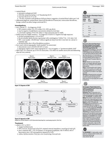
Blood in basal cisterns
Blood in suprasellar cistern
Blood in interhemispheric fissure
Nontraumatic Subarachnoid Hemorrhage in the Setting of Negative Cranial Computed Tomography Results: External Validation of a Clinical and Imaging Prediction Rule
Ann Emerg Med 2012;59:460-8.e1-7.doi:10.1016 Background: Two rules for SAH diagnosis exist. A clinical prediction rule states that patients with acute severe H/A but without the clinical variables age ≥40 yr, neck pain, loss of consciousness, or onset of H/A with exertion are at low risk for SAH. An imaging prediction rule bases diagnosis on non-contrast cranial CT for patients within 6 h of H/A onset.
Methods: Matched case-control study of 55 patients at 21 emergency departments between 2000 and 2011, and diagnoses were verified by lumbar puncture.
Results: The clinical prediction rule for diagnosis of SAH was 97.1% sensitive, 22.7% specific, and had a negative likelihood ratio of 0.13. Using the imaging prediction rule resulted in a false negative rate of 20%.
Conclusions: Performing the clinical and imaging rules together has the potential for maximizing sensitivity of prediction and reducing rates of lumbar puncture, but using imaging alone can result in missed cases.
The Vasograde: A Simple Grading Scale For Prediction Of Delayed Cerebral Ischemia After Subarachnoid Hemorrhage
Stroke 2015; 46(7):1826-31
Background: Patients are classically at risk of delayed cerebral ischemia (DCI) after aneurysmal subarachnoid hemorrhage. We validated a grading scale—the VASOGRADE—for prediction of DCI. Method: We used data of 3 phase II randomized clinical trials and a single hospital series to assess the relationship between the VASOGRADE and DCI. Results: In a cohort of 746 patients, the VASOGRADE signicantly predicted DCI (P<0.001). The VASOGRADE-Yellow had a tendency for increased risk for DCI (odds ratio [OR], 1.31; 95% CI, 0.77–2.23) when compared with VASOGRADE- Green; those with VASOGRADE-Red had a 3-fold higher risk of DCI (OR, 3.19; 95% CI, 2.07–4.50). VASOGRADE had an adequate discrimination for prediction of DCI (area under the receiver operating characteristics curve=0.63) and good calibration. Conclusions: The VASOGRADE results validated previously published risk charts in a large and diverse sample of subarachnoid hemorrhage patients, which allows DCI risk stratification on presentation after subarachnoid hemorrhage. It could help to select patients at high risk of DCI,
as well as standardize treatment protocols and research studies.
The VASOGRADE: A simple grading scale for prediction of delayed cerebral ischemia after subarachnoid hemorrhage
Stroke 2015;46: 1826-1831
Blood on surface of tentorium
Blood in Sylvian fissures
CTA
Positive
Figure 19. Diagnosis of SAH
MRI/MRA
Stop
NCCT
LP
Stop Gold Standard
CTA
Stop
Positive
CT or MRA positive
LP positive Diagnostic DSA
Yes
Aneurysm Treatment
OR DSA
No
Diagnostic DSA Gold Standard
Figure 20. Approach to SAH
Adapted from: de Oliveira Manoel et al. (2014) Subarachnoid haemorrhage from a neuroimaging perspective. Critical Care
Treatment
• admittoICUorNICU
■ oxygen/ventilationprn
■ NPO, bed rest, elevate head of bed 30o, minimal external stimulation, neurological vitals q1h ■ aim to maintain sBP = 120-150 (balance of vasospasm prophylaxis, risk of rebleed, risk of
hypotension since CBF autoregulation impaired by SAH)
■ cardiac rhythm monitor, Foley prn, strict monitoring of ins and outs
(Clipping)
(Coiling)
Negative
Negative Negative
Negative
