
Page 832 - TNFlipTest
P. 832
NS34 Neurosurgery
Neurotrauma Toronto Notes 2019
Medical Management Specific to Spinal Cord Injury
• option:methylprednisolone(givenwithin8hofinjury)thisiscontroversialandyouneedtoconfer with Neurosurgery service
• ±decompressioninacute,non-penetratingspinalcordinjury
Fractures of the Spine
FRACTURES AND FRACTURE-DISLOCATIONS OF THE THORACIC AND LUMBAR SPINE
• assessligamentousinstabilityusingflexion/extensionx-rayviewsof±MRI
• thoracolumbarspineunstableif4/6segmentsdisrupted(3columnsdividedintoleftandright)
■ anterior column: anterior half of vertebral body, disc, and anterior longitudinal ligament
■ middle column: posterior half of vertebral body, disc, and posterior longitudinal ligament
■ posterior column: posterior arch, facet joints, pedicle, lamina and supraspinous, interspinous, and
ligamentum ligaments
Types of Injury
Table 20. Denis Classification of Spinal Trauma
Fracture Type
Compression Fracture (58%)
Burst Fracture (17%)
Flexion Distraction Injury (6%)
Fracture-Dislocation (6%)
Description
Produced by flexion
Posterior ligament complex (supraspinous and interspinous ligaments, ligamentum flavum, and intervertebral joint capsules) remain intact
Fractures are stable but lead to kyphotic deformity
Stable: anterior and middle columns parted with bone retropulsed nearby
Hallmark is pedicle widening on AP x-ray
Spinal cord (seen on x-ray and CT); posterior column is uninjured
Unstable: same as the stable but with posterior column disruption (usually ligamentous)
Hyperflexion and distraction of posterior elements
Middle and posterior columns fail in distraction
Classic: Chance, horizontal fracture through posterior arch, pedicles, posterior vertebral body Can be purely ligamentous, i.e. through PLL and disc
Anterior and cranial dislocation of superior vertebral body → 3 column failure
Three types: (1) flexion-rotation, (2) flexion-distraction, (3) shear/hyperextension (rare)
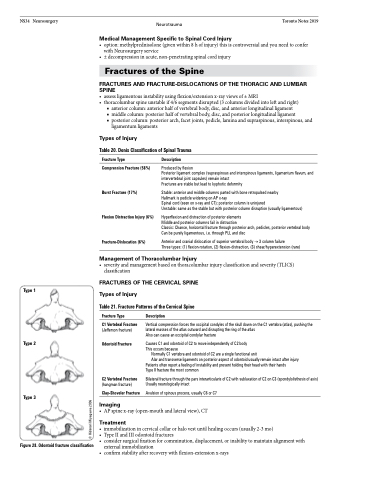
Type 1
Type 2
Type 3
Figure 28. Odontoid fracture classification
Management of Thoracolumbar Injury
• severityandmanagementbasedonthoracolumbarinjuryclassificationandseverity(TLICS) classification
FRACTURES OF THE CERVICAL SPINE Types of Injury
Table 21. Fracture Patterns of the Cervical Spine
Fracture Type
C1 Vertebral Fracture
(Jefferson fracture)
Odontoid Fracture
C2 Vertebral Fracture
(hangman fracture)
Clay-Shoveler Fracture
Imaging
Description
Vertical compression forces the occipital condyles of the skull down on the C1 vertebra (atlas), pushing the lateral masses of the atlas outward and disrupting the ring of the atlas
Also can cause an occipital condylar fracture
Causes C1 and odontoid of C2 to move independently of C2 body This occurs because
Normally C1 vertebra and odontoid of C2 are a single functional unit
Alar and transverse ligaments on posterior aspect of odontoid usually remain intact after injury Patients often report a feeling of instability and present holding their head with their hands
Type II fracture the most common
Bilateral fracture through the pars interarticularis of C2 with subluxation of C2 on C3 (spondylolisthesis of axis) Usually neurologically intact
Avulsion of spinous process, usually C6 or C7
• APspinex-ray(open-mouthandlateralview),CT
Treatment
• immobilizationincervicalcollarorhalovestuntilhealingoccurs(usually2-3mo)
• TypeIIandIIIodontoidfractures
• consider surgical fixation for comminution, displacement, or inability to maintain alignment with
external immobilization
• confirmstabilityafterrecoverywithflexion-extensionx-rays
© Hidenori Miyagawa 2006
