
Page 830 - TNFlipTest
P. 830
NS32 Neurosurgery
Neurotrauma Toronto Notes 2019
• basalskullfractures:notreadilyseenonx-ray,relyonclinicalsigns ■ retroauricular ecchymoses (Battle’s sign)
■ periorbital ecchymoses (raccoon eyes)
■ hemotympanum
■ CSF rhinorrhea, otorrhea (suspect CSF if halo or target sign present); suspect with Lefort II/III midface fracture
Cranial Nerve Injury
• mosttraumaticcausesofcranialnerveinjurydonotwarrantsurgicalintervention • surgicalintervention
■ CN II: local eye/orbit injury
■ CN III, IV, VI: if herniation secondary to mass ■ CN VIII: repair of ossicles
• CNinjuriesthatimprove
■ CN I: recovery may occur in a few months; most do not improve
■ CN III, IV, VI: majority recover
■ CN VII: recovery with delayed lesions
■ CN VIII: vestibular symptoms improve over weeks, deafness usually permanent (except when
resulting from hemotympanum)
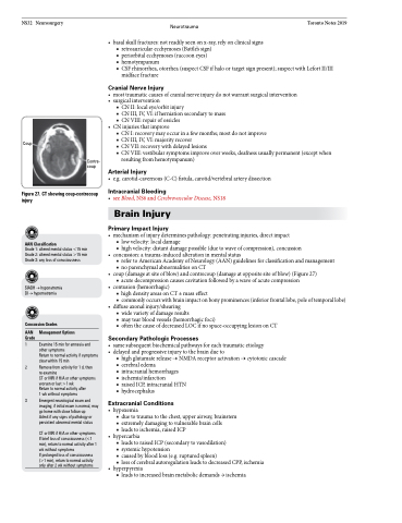
Coup
Figure 27. CT showing coup-contrecoup injury
AAN Classification
Grade 1: altered mental status <15 min Grade 2: altered mental status >15 min Grade 3: any loss of consciousness
SIADH → hyponatremia DI → hypernatremia
Concussion Grades
AAN Management Options Grade
1 Examine 15 min for amnesia and other symptoms
Return to normal activity if symptoms
clear within 15 min
2 Remove from activity for 1 d, then
re-examine
CT or MRI if H/A or other symptoms worsen or last >1 wk
Return to normal activity after
1 wk without symptoms
3 Emergent neurological exam and imaging; if initial exam is normal, may go home with close follow-up
Admit if any signs of pathology or persistent abnormal mental status
CT or MRI if H/A or other symptoms If brief loss of consciousness (<1 min), return to normal activity after 1 wk without symptoms
If prolonged loss of consciousness (>1 min), return to normal activity only after 2 wk without symptoms
Contre- coup
Arterial Injury
• e.g.carotid-cavernous(C-C)fistula,carotid/vertebralarterydissection
Intracranial Bleeding
• seeBlood,NS6andCerebrovascularDisease,NS18 Brain Injury
Primary Impact Injury
• mechanismofinjurydeterminespathology:penetratinginjuries,directimpact
■ low velocity: local damage
■ high velocity: distant damage possible (due to wave of compression), concussion
• concussion:atrauma-inducedalterationinmentalstatus
■ refer to American Academy of Neurology (AAN) guidelines for classification and management ■ no parenchymal abnormalities on CT
• coup(damageatsiteofblow)andcontrecoup(damageatoppositesiteofblow)(Figure27) ■ acute decompression causes cavitation followed by a wave of acute compression
• contusion(hemorrhagic)
■ high density areas on CT ± mass effect
■ commonlyoccurswithbrainimpactonbonyprominences(inferiorfrontallobe,poleoftemporallobe)
• diffuseaxonalinjury/shearing
■ wide variety of damage results
■ may tear blood vessels (hemorrhagic foci)
■ often the cause of decreased LOC if no space-occupying lesion on CT
Secondary Pathologic Processes
• samesubsequentbiochemicalpathwaysforeachtraumaticetiology • delayedandprogressiveinjurytothebraindueto
■ highglutamaterelease→NMDAreceptoractivation→cytotoxiccascade ■ cerebral edema
■ intracranial hemorrhages
■ ischemia/infarction
■ raised ICP, intracranial HTN ■ hydrocephalus
Extracranial Conditions
• hypoxemia
■ due to trauma to the chest, upper airway, brainstem ■ extremely damaging to vulnerable brain cells
■ leads to ischemia, raised ICP
• hypercarbia
■ leads to raised ICP (secondary to vasodilation)
■ systemic hypotension
■ caused by blood loss (e.g. ruptured spleen)
■ loss of cerebral autoregulation leads to decreased CPP, ischemia
• hyperpyrexia
■ leads to increased brain metabolic demands → ischemia
