
Page 824 - TNFlipTest
P. 824
NS26 Neurosurgery
Cervical spondylotic myelopathy is the most common cause of spinal cord impairment
Clinical Grading Scores to Assess CSM
• mJOA
• Nurick Grade
• Neck Disability Index
A Clinical Practice Guideline for the Management of Patients with Degenerative Cervical Myelopathy (Dcm): Recommendations for Patients with Mild, Moderate, and Severe Disease and Nonmyelopathic Patients with Evidence of Cord Compression
Global Spine Journal 2017; 7(3S):70S-83S.
Severe and moderate DCM: moderate evidence suggesting strong recommendation of surgical intervention.
Mild DCM: very low to low evidence suggesting offering surgical intervention or a structured rehabilitation and if non-operative management initially pursued, consider operative intervention if evidence of neurological deterioration. Non-myelopathic patients without radiculopathy: in such patients with imaging evidence of cervical cord compression, suggestion of not offering prophylactic surgery; counsel, educate, and follow clinically.
Non-myelopathic patients with radiculopathy: such patients with imaging evidence of cervical cord compression are at a higher risk of developing myelopathy and should be counselled. Offer surgical or nonoperative treatment with appropriate follow- up and structured rehabilitation.
Extradural Lesions Toronto Notes 2019 Cervical Spondylosis
Definition
• progressivedegenerativeprocessofcervicalspineleadingtocanalstenosis–congenitalspinalstenosis, degeneration of intervertebral discs, hypertrophy of lamina, dura, or ligaments, subluxation, altered mobility, telescoping of the spine due to loss of height of vertebral bodies, alteration of normal lordotic curvature
• resultantsyndromes:mechanicalneckpain,radiculopathy(rootcompression),myelopathy(spinalcord compression)
Epidemiology
• typicallybeginsatage40-50,M>F,mostcommonlyattheC5-C6>C6-C7levels
Pathogenesis
• anyof:discdegeneration/herniation,osteophyteformation,ossification,andhypertrophyofligaments • pathophysiologyincludesstaticcompression,dynamiccompression,andvascularcompromise
Clinical Features
• insidiousonsetofmechanicalneckpainexacerbatedbyexcessvertebralmotion(particularlyrotation
• • •
•
and lateral bending with a vertical compressive force – Spurling’s test) theearliestsymptomsaregaitdisturbanceandlowerextremityweaknessorstiffness occipitalH/Aiscommon radiculopathymayinvolve1ormoreroots,andsymptomsincludeneck,shoulder,andarmpain, paresthesias and numbness
cervicalspondyloticmyelopathymaypresentwith:
■ weakness (upper > lower extremity), lower extremity weakness (corticospinal tracts) is most
worrisome complaint
■ decreased dexterity, loss of fine motor control
■ sensory changes
■ UMN findings such as hyperreflexia, clonus, and Babinski reflex
■ funicular pain, characterized by burning and stinging ± Lhermitte’s sign (lightning-like sensation
down the back with neck flexion)
Investigations
• x-ray of cervical spine ± flexion/extension (alignment, fractures)
• MRImostusefulfordeterminationofcompressionoftheneuralelement
• CTisonlyusedforbetterdeterminationofbonyanatomy(i.e.OPLL)
• EMG/nerveconductionstudiesreservedforperipheralnerveinvestigation
Treatment
• nonsurgical:physiotherapy,anti-inflammatorymedications
• surgical:anteriorapproach(anteriorcervicaldiscectomyorcorpectomy),posteriorapproach
(decompressive cervical laminectomy)
• in multilevel CSM, an anterior approach is associated with better postoperative neural function but has
a higher complication and reoperation rate than the posterior group
• surgicalindications:myelopathywithmotorimpairment,progressiveneurologicimpairment,
intractable pain
• completeremissionalmostneveroccurs;surgicaldecompressionmaystopprogressionofdisease
Lumbar Disc Syndrome
Etiology
• posteriolaterallyherniateddisccompressednerverootexitingBELOWthelevelofthediscorthe traversing nerve root
• farlateraldischerniationcompressednerverootATthelevelofthediscortheexitingnerveroot
• centralherniationcausescaudaequinaorlumbarstenosis(neurogenicclaudication)
Clinical Features
• initiallybackpain,thenlegpain>backpain
• limitedbackmovement(especiallyforwardflexion)duetopain
• motorweakness,dermatomalsensorychanges,decreasedreflexes • exacerbationwithvalsalva;reliefwithflexingthekneeorthigh
• nerveroottensionsigns
■ straight leg raise (Lasegue’s test) or crossed SLR (pain should occur at less than 60o) suggests L5, S1 root involvement
■ femoral stretch test suggests L2, L3, or L4 root involvement
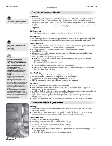
Figure 23. T2 weighted MRI of lumbar disc herniation
