Page 825 - TNFlipTest
P. 825
 Toronto Notes 2019 Extradural Lesions
Investigations
• MRIismodalityofchoice
• x-rayspine(onlytoruleoutotherlesions),CT(bonyanatomy)
• myelogramandpost-myelogramCT(onlyifMRIiscontraindicated)
Treatment
• conservative(sameascervicaldiscdisease)
• surgicalindications:sameascervicaldiscandcaudaequinasyndrome
Prognosis
• 95%improvespontaneouslywithin4-8wk
• thosewhodonotimprovewithconservativetreatmentachievesymptomreliefquickerwithsurgery
than continuation of conservative management; however, the long-term outcome after surgery is
comparable to conservative therapy
• do not follow patients with serial MRIs; clinical status is more important at guiding management
Neurosurgery NS27
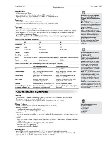
          Table 15. Lateral Lumbar Disc Syndromes
Surgical versus Non-Operative Treatment for Lumbar Disc Herniation: Eight-Year Results for the Spine Patient Outcomes Research Trial (SPORT)
Spine 2014; 39(1): 3–16.
Methods: In the RCT arm of the study, 501 patients with imaging-confirmed lumbar disc herniation were randomized to open discectomy versus standard non-operative management.
Results: Surgery was superior in intent-to-treat analysis for sciatica severity (p=0.005), patient satisfaction (p=0.013), and self-rated improvement (p=0.013) at 8 year follow-up. Improvements in pain, physical function, and disability were only seen in an as-treated analysis due to significant non-adherence to treatment assignment. Conclusion: Patients who are carefully selected
for surgical intervention show greater symptom improvement compared to non-operative management.
Magnetic Resonance Imaging in Follow-Up Assessment of Sciatica
NEJM 2013;368:999-1007
Background: Follow-up MRI is a controversial method for monitoring sciatica in patients with known lumbar-disc herniation.
Methods: Participants (n=283) were recruited from a simultaneous, parallel, randomized study comparing surgery and conservative care for sciatica (the Sciatica Trial). MRI and clinical assessment were undertaken pre-treatment and 1 yr post-treatment randomization to visualize disc herniation and evaluate outcome.
Results: At 1 yr, disc herniation was visible in 35% with a favourable outcome (complete, or nearly complete symptom resolution) and in 33% with
an unfavourable outcome (p=0.70). A favourable outcome was reported in 85% of patients with disc herniation and 83% without disc herniation (p=0.70).
Conclusions: Anatomical abnormalities visible on repeated MRI 1 yr after treatment for sciatica due to lumbar-disc herniation could not distinguish patients with resolution of their symptoms from patients still experiencing symptoms.
Causes of Cauda Equina Syndrome
• Lumbar disc herniation • Spinal stenosis
• Spinal tumour
• Epidural abscess
• Hematoma • Trauma
  Root Involved Incidence Pain
Sensory Lateral leg Motor
Reflex
L3-4
L4
<10%
Femoral pattern
Medial leg
Lateral foot
Tibialis anterior (dorsiflexion) Patellar
L4-5
L5
45%
Sciatic pattern Dorsal foot to hallux
Extensor hallucis longus (hallux extension) Medial hamstrings
L5-S1
S1
45%
Sciatic pattern
Gastrocnemius, soleus (plantar flexion) Achilles
           Table 16. Differentiating Conus Medullaris Syndrome from Cauda Equina Syndrome
  Onset Spontaneous Pain
Sensory Deficit Motor Deficit
Reflexes
Autonomic Symptoms (bladder dysfunction, impotence, etc.)
Conus Medullaris Syndrome
Sudden, bilateral
Rare, if present usually bilateral, symmetric in perineum or thighs
Saddle; bilateral and symmetric; sensory dissociation
Symmetric; paresis less marked; fasciculations may be present
Only ankle jerk absent (preserved knee jerk)
Urinary retention and atonic anal sphincter prominent early; impotence frequent
Cauda Equina Syndrome
Gradual, unilateral
Severe, radicular type: in perineum, thighs, legs, back, or bladder
Saddle; no sensory dissociation; may be unilateral and asymmetric
Asymmetric; paresis more marked; atrophy may be present; fasciculations rare
Knee and ankle jerk may be absent
Sphincter dysfunction presents late; impotence less frequent
  Cauda Equina Syndrome
Etiology
• compressionorirritationoflumbosacralnerverootsbelowconusmedullaris(belowL2level) • decreasedspaceinthevertebralcanalbelowL2
• commoncauses:herniateddisc±spinalstenosis,vertebralfracture,andtumour
Clinical Features
• usuallyacute(developsinlessthan24h);rarelysubacuteorchronic • motor(LMNsigns)
■ weakness in multiple root distribution
■ reduced deep tendon reflexes (knee or ankle) • autonomic
■ urinary retention (or overflow incontinence) and/or fecal incontinence due to loss of anal sphincter tone
• sensory
■ low back pain radiating to legs (sciatica) aggravated by Valsalva maneuver and by sitting; relieved by
lying down
■ bilateral sensory loss or pain: depends on the level affected
■ saddle area (S2-S5) anesthesia
■ sexual dysfunction (late finding)
Investigations
• urgentMRItoconfirmcompressionofS2-S3-S4nerverootbyalargedischerniation
• post-voidresidualveryhelpfultodetermineiftrueretentionispresent;volumescontroversialbut
  anything over 250 cc in a healthy individual is cause for concerns


