
Page 822 - TNFlipTest
P. 822
NS24 Neurosurgery
Cerebrospinal Fluid Fistulas
Toronto Notes 2019
Suspect CSF fistula in patients with otorrhea or rhinorrhea after head trauma or recurrent meningitis
Cerebrospinal Fluid Fistulas
Etiology
• cranialorspinal
• traumatic:afterheadtrauma,iatrogenic(post-transsphenoidalsurgery,postskullbasesurgery)
• nontraumatic:highpressure(hydrocephalus,tumour),normalpressure(boneerosionsecondaryto
infection, congenital defect)
Clinical Features
• otorrheaorrhinorrhea(clearfluid)
• lowpressureheadaches(worsewhensittingup)
• confirmatorytestingforCSF:βtransferrintest,quantitativeglucoseanalysisoffluid,“ringsign”,
“reservoir sign”
Investigations
• CT(detectpneumocephalus,fractures,skullbasedefects),watercontrastCTcisternography
Treatment
• lowerICP(avoidstraining,acetazolamidetoreduceCSFproduction,modestfluidrestriction)
Ring Sign: If CSF is stringed with blood. Allow CSF to drain onto the surrounding sheets; positive if clear in centre with surrounding blood coloured ring (double ring sign) Reservoir sign: Gush of CSF leaks out in certain head positions; i.e. teapot sign (not specific or sensitive)
Stereotactic Radiosurgery For Cavernous Malformations (CM)
J Neurosurg. 2010;113(4):691-9
Summary: stereotactic radiosurgery is a safe intervention for CMs, with advantages of reducing rebleed risks in patients with repeated pretreatment hemorrhage. Treating CMs with single bleed is
less clear. However, the morbidity of repeated hemorrhage outweights any radiosurgery related morbidity and early active management of deep- seated CMs should be considered.
Methods: retrospective analysis of 113 patients with 79 brainstem and 39 thalamic/basal ganglia CMs treated with gamma knife surgery. Results:
1. Patientswithmultiplesymptomatichemorrhages before radiosurgery (n=41): rebleed rate decreased from 30.5% per lesion to 15% for
the first 2 years after radiosurgery and 2.4% subsequently. Pretreatment multiple bleeds led to permanent deficits in 72% of these patients.
2. Patientswith≤1symptomaticbleedbefore radiosurgery (n=77): natural history is uncertain due to short period between presenting
bleed and treatment (median 1 year). Rate of hemorrhage was 5.1% for the first 2 years and 1.3% subsequently. Pretreatment hemorrhages led to permanent deficits in 43% of these patients (significantly lower than multiple-bleeds group, p<0.001)
3. Permanent adverse radiation effects were rare (7.3%) and minor in both groups.
4. Posttreatment hemorrhages led to persistent deficits in 7.3% of patients.
RED FLAGS for Back Pain
BACK PAIN
Bowel/Bladder (retention or incontinence) Anesthesia (saddle)
Constitutional symptoms “K”hronic disease Parasthesia
Age >50 yr or <20 yr IV drug use
Neuromotor deficits
Cauda Equina
• Urinaryretentionorincontinence,fecal
incontinence or loss of anal sphincter tone, saddle
anesthesia, uni/bilateral, leg weakness/pain
Malignancy
• Age>50yr,previousHxofcancer,pain unrelieved by bed rest, constitutional symptoms
Infection
• IncreasedESR,IVdruguse,immunosuppressed, fever
Compression Fracture
• Age>50yr,trauma,prolongedsteroiduse
• •
•
persistentleak:mayrequirecontinuouslumbardrainageviapercutaneouscatheter surgicalindications:traumaticleaklasting>2wk,spontaneousleaks,delayedonsetofleakaftertrauma or surgery, leaks complicated by meningitis
EXTRACRANIAL PATHOLOGY Approach to Limb/Back Pain
seeOrthopedics,OR5
Extradural Lesions
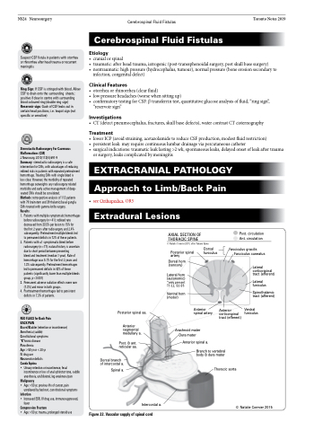
AXIAL SECTION OF
THORACIC SPINE
© Natalie Cormier 2015, after Takami Iijima
Post. circulation Ant. circulation
Fasciculus gracilis Fasciculus cuneatus
Posterior spinal artery
Dorsal horn (sensory)
Lateral horn (autonomic) *only present T1-L2, S2-S5
Ventral horn (motor)
Dorsal funiculus
Posterior spinal aa.
Anterior spinal artery
Anterior corticospinal tract (efferent)
Lateral corticospinal tract (efferent)
Lateral funiculus
Spinothalamic tract (afferent)
Ventral funiculus
Anterior segmental medullary a.
Post. & ant. reticular aa.
Dorsal branch of intercostal a.
Spinal a.
Arachnoid mater Dura mater
Anterior spinal a.
Branch to vertebral body & dura mater
Thoracic aorta
Figure 22. Vascular supply of spinal cord
Intercostal a.
© Natalie Cormier 2015
