
Page 897 - TNFlipTest
P. 897
Toronto Notes 2019
Optics
Ophthalmology OP7
SLIT-LAMP EXAMINATION Ocular Adnexa
• lids, lashes, lacrimal system
Anterior Segment
• conjunctiva/sclera • cornea
■ fluorescein dye: stains de-epithelialized cornea; dye appears fluorescent green with cobalt blue filtered light
■ Rose Bengal dye: stains devitalized corneal epithelium • anteriorchamber/angle(VanHerick)
• iris/pupil
• lens(assessforcataract)
• anteriorvitreous
Posterior Segment (requires 78D or 90D lens)
• vitreous
• opticdisc(colour,C:Dratio,sharpnessofdiscmargin)
• macula(~1.5-2discdiameterstemporaltodisc),fovea(foveallightreflex) • retinalvessels
• retinalbackground
TONOMETRY
• measurementofIOP
• normalrangeis9-21mmHg(average15mmHg)
• IOPhasdiurnalvariation,soalwaysrecordthetimeofdayatwhichthemeasurementwastaken • commonlymeasuredby:
■ GAT: clinical gold standard, performed using the slit-lamp with special tip (prism)
■ Tono-Pen®: benefit is portability and use of disposable probe tips; use when GAT is inaccurate, such
as when the cornea is scarred or asymmetric ■ air puff (non-contact and least reliable)
• usetopicalanestheticforGATandTono-Pen®;applyfluoresceindyeandusecobaltbluelightforGAT
DIRECT OPHTHALMOSCOPY
• bestperformedwithpupilsdilated(forlistofmydriaticsandcycloplegicsseeTable13,OP42) 1. assess red reflex
◆ light reflected off the retina produces a “red reflex” when viewed from ~1 foot away
◆ anything that interferes with the passage of light will diminish the red reflex (e.g. large vitreous
hemorrhage, cataract, retinoblastoma) 2. examine the posterior segment of the eye
◆ vitreous
◆ optic disc (colour, C:D ratio, sharpness of disc margin)
◆ macula (~1.5-2 disc diameters temporal to disc), fovea (foveal light reflex) ◆ retinal vessels
◆ retinal background
• contraindicationstopupillarydilatation
■ shallow anterior chamber – can precipitate acute angle-closure glaucoma
■ iris-supported anterior chamber lens implant
■ potential neurologic abnormality requiring pupil evaluation
■ use caution with cardiovascular disease – mydriatics may cause tachycardia and HTN
Optics
REFRACTION
• twotechniquesused
■ flash/streak retinoscopy: refractive error determined objectively by the examiner using lenses and
retinoscope
■ manifest: subjective trial using loose lenses or a phoropter (device the patient looks through that is
equipped with lenses)
■ cycloplegic: manifest refraction with accommodation temporarily paralyzed with cycloplegics
• atypicallensprescriptionwouldcontain:
■ sphere power in dioptre (measurement of refractive power of lens, equal to reciprocal of focal length
in metres)
■ cylinder power in dioptre to correct astigmatism
■ axis of cylinder in degrees
■ “add” (bifocal/progressive reading lens) for presbyopes
■ e.g. -1.50 + 1.00 x 120o, add +2.00
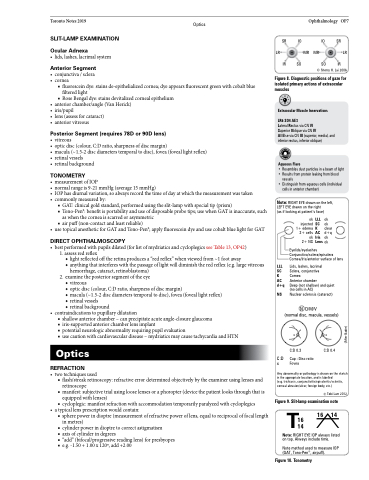
LR
SR IO IO SR MR MR LR
IR SO SO IR
© Sherry H. Lai 2006
Figure 8. Diagnostic positions of gaze for isolated primary actions of extraocular muscles
Extraocular Muscle Innervations
LR6 SO4 AE3
Lateral Rectus via CN VI
Superior Oblique via CN IV
All Else via CN III (superior, medial, and inferior rectus, inferior oblique)
AqueousFlare
• Resembles dust particles in a beam of light
• Results from protein leaking from blood
vessels
• Distinguish from aqueous cells (individual
cells in anterior chamber)
Note: RIGHT EYE drawn on the left, LEFT EYE drawn on the right
(as if looking at patient’s face)
LLL SC K AC d+q
NS
C:D x
Eyelids/eyelashes Conjunctiva/sclera/episclera Cornea/Iris/anterior surface of lens
Lids, lashes, lacrimal
Sclera, conjunctiva
Cornea
Anterior chamber
Deep (not shallow) and quiet (no cells in AC)
Nuclear sclerosis (cataract)
N D/M/V
(normal disc, macula, vessels)
xx
ok injected 1+ edema 2+ cells ok 2+ NS
LLL ok SC ok
K clear AC d+q Iris ok Lens ok
C:D 0.3
Cup : Disc ratio Fovea
C:D 0.4
Any abnormality or pathology is drawn on the sketch in the appropriate location, and is labelled
(e.g. trichiasis, conjunctivitis/episcleritis/scleritis, corneal abrasion/ulcer, foreign body, etc.)
© Tobi Lam 2012
Figure 9. Slit-lamp examination note 16 16 14
T
14
Note: RIGHT EYE lOP always listed
on top. Always include time.
Note method used to measure lOP (GAT, Tono-Pen®, airpuff).
Figure 10. Tonometry
(After Bader)
