
Page 843 - TNFlipTest
P. 843
Toronto Notes 2019 Maternal Physiologic Adaptations to Pregnancy
■ Parity (TPAL)
◆ T: number of term deliveries (>37 wk)
◆ P: number of premature deliveries (20-36+6 wk) ◆ A: number of abortions (ending <20 wk)
– induced (therapeutic) and spontaneous (miscarriage) ◆ L: number of living children
Physical Signs
• uterineenlargement
• breastengorgement,areoladarkening,andprominentvascularpatterns
• Goodell’ssign:softeningofthecervix(4-6wk)
• Chadwick’ssign:bluishdiscolourationofthecervixandvaginaduetopelvicvasculatureengorgement
(6 wk)
• Hegar’ssign:softeningofthecervicalisthmus(6-8wk)
Investigations
• β-hCG:peptidehormonecomposedofαandβsubunitsproducedbyplacentaltrophoblasticcells– maintains the corpus luteum during pregnancy
■ positive in serum 9 d post-conception, positive in urine 28 d after first day of LMP
■ plasma levels usually double every 1.4-2.0 d, peak at 8-12 wk, then fall, but continue to be
measurable until delivery
• levelslessthanexpectedsuggest:ectopicpregnancy,abortion,inaccuratedates,andsomenormal
pregnancies
• levelsgreaterthanexpectedsuggest:multiplegestation,molarpregnancy,Trisomy21,orinaccurate
dates
• U/S:
■ transvaginal
◆ 5 wk GA: gestational sac visible ◆ 6 wk: fetal pole visible
◆ 7-8 wk: fetal heart activity visible
■ transabdominal
◆ 6-8 wk: intrauterine pregnancy visible
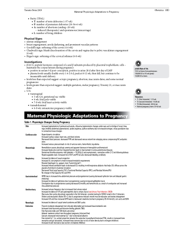
Maternal Physiologic Adaptations to Pregnancy
Table 1. Physiologic Changes During Pregnancy
Obstetrics OB3
β-hCG Rule of 10s
10 IU at time of missed menses 100,000 IU at 10 wk (peak) 10,000 IU at term
Trimesters
• T1 (first trimester): 1-14 wk
• T2 (second trimester): 14-28 wk • T3 (third trimester): 28-42 wk
• Normal pregnancy term: 37-42 wk
Skin Cardiovascular
Hematologic
Respiratory
Gastrointestinal
Genitourinary
Neurologic Endocrine
Increased pigmentation of perineum and areola, chloasma (pigmentation changes under eyes and on bridge of nose), linea nigra (midline abdominal pigmentation), spider angiomas, palmar erythema due to increased estrogen, striae gravidarum due to connective tissue changes
Hyper-dynamic circulation
Increased cardiac output, heart rate, and blood volume
Decreased blood pressure: decreased PVR and decreased venous return from enlarging uterus compressing IVC and pelvic veins
Increased venous pressure leads to risk of varicose veins, hemorrhoids, leg edema
Hemodilution causes physiologic anemia and apparent decrease in hemoglobin and hematocrit
Increased leukocyte count but impaired function leads to improvement in some autoimmune diseases
Gestational thrombocytopenia: mild (platelets >70,000/μL) and asymptomatic, normalizes within 2-12 wk following delivery Hypercoagulable state: increased risk of DVT and PE but also decreased bleeding at delivery
Increased incidence of nasal congestion
Increased O2 consumption to meet increased metabolic requirements
Elevated diaphragm (i.e. appears more “barrel-chested”)
Increased minute ventilation leads to decreased CO2 resulting in mild respiratory alkalosis that helps CO2 diffuse across the placenta from fetal to maternal circulation
Decreased Total Lung Capacity (TLC), Functional Residual Capacity (FRC), and Residual Volume (RV)
No change in Vital Capacity (VC) and FEV1
GERD due to increased intra-abdominal pressure and progesterone (causing decreased sphincter tone and delayed gastric emptying)
Increased incidence of gallstones due to progesterone causing increased gallbladder stasis
Constipation due to progesterone causing decreased GI motility and hemorrhoids as a result of constipation and increased intra-abdominal pressure
Increased urinary frequency due to increased total urinary output
Increased incidence of UTI and pyelonephritis due to urinary stasis (see Urinary Tract Infection, OB29)
Glycosuria that can be physiologic especially in the 3rd trimester; consider testing for GDM if noted in first 2 trimesters Ureters and renal pelvis dilation (R>L) due to progesterone-induced smooth muscle relaxation and uterine enlargement Increased CO and thus increased GFR leads to decreased creatinine (normal in pregnancy 35-44 mmol/L), uric acid, and BUN
Increased incidence of carpal tunnel syndrome and Bell’s palsy
Thyroid: moderate enlargement (not clinically detectable) and increased basal metabolic rate Increased total thyroxine and thyroxine binding globulin (TBG)
Free thyroxine index and TSH levels are normal
Adrenal: maternal cortisol rises throughout pregnancy (total and free)
Calcium: decreased total maternal Ca2+ due to decreased albumin
Free ionized Ca2+ (i.e. active) proportion remains the same due to parathyroid hormone (PTH), results in increased bone resorption and gut absorption, increased bone turnover (but no loss of bone density due to estrogen inhibition) Increased insulin resistance (see section on Diabetes Mellitus)
