
Page 800 - TNFlipTest
P. 800
NS2 Neurosurgery
Acronyms
Toronto Notes 2019
AVF AVM BBB CBF CNS CPA CPP CSF CVR DBS DI ECF ECT EEG
arteriovenous fistula arteriovenous malformation blood brain barrier
cerebral blood flow
central nervous system cerebellar pontine angle cerebral perfusion pressure cerebral spinal fluid cerebral vascular resistance deep brain stimulation diabetes insipidus extracellular fluid electroconvulsive therapy electroencephalography
See Functional Neuroanatomy Software
EMG EVD GCS GPi H/A IC ICF ICH ICP ICU IVH LMN LOC
electromyography
external ventricular drain Glasgow coma scale globus pallidus pars interna headache
internal capsule intracellular fluid intracerebral hemorrhage intracranial pressure intensive care unit intraventricular hemorrhage lower motor neuron
loss of consciousness
LP lumbar puncture SAH MAP mean arterial pressure SCI MLS midline shift SDH N/V nausea/vomitting SIADH NC neurogenic claudication
NICU neonatal intensive care unit SPECT
subarachnoid hemorrhage
spinal cord injury
subdural hemorrhage
syndrome of inappropriate antidiuretic hormone
single photon emission computed tomography
stereotactic radiosurgery
Acronyms
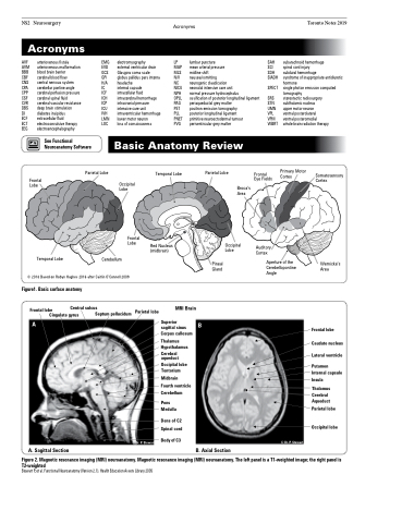
Frontal Lobe
Occipital Lobe
Frontal Lobe
Broca’s Area
Temporal Lobe
© 2018 Based on Robyn Hughes 2016 after Caitlin O’Connell 2009
Figure1. Basic surface anatomy
Frontal lobe Central sulcus
A
Superior sagittal sinus Corpus callosum
Thalamus
Hypothalamus
Cerebral aqueduct
Occipital lobe
Tentorium
Midbrain Fourth ventricle Cerebellum
Pons Medulla
Dens of C2 Spinal cord
Body of C3
B
A. Sagittal Section
B. Axial Section
Cingulate gyrus
Septum pellucidum
Parietal Lobe
Temporal Lobe
Parietal Lobe
Frontal Eye Fields
Auditory Cortex
Primary Motor Cortex
Somatosensory Cortex
Cerebellum
Pineal Gland
Aperture of the Cerebellopontine Angle
Wernicke’s Area
Frontal lobe
Caudate nucleus Lateral ventricle
Putamen Internal capsule Insula
Thalamus Cerebral Aqueduct
Parietal lobe Occipital lobe
Parietal lobe
MRI Brain
© Dr. P. Stewart
© Dr. P. Stewart
Red Nucleus (midbrain)
Occipital Lobe
NPH normal pressure hydrocephalus
OPLL ossification of posterior longitudinal ligament PAG periaqueductal grey matter
PET positron emission tomography
PLL posterior longitudinal ligament
PNET primitive neuroectodermal tumour
PVG periventricular grey matter
SRS
STN subthalamic nucleus UMN upper motor neuron
VPL ventral posterolateral
VPM ventral posteromedial
WBRT whole brain radiation therapy
Basic Anatomy Review
Figure 2. Magnetic resonance imaging (MRI) neuroanatomy. Magnetic resonance imaging (MRI) neuroanatomy. The left panel is a T1-weighted image; the right panel is
T2-weighted
Stewart P, et al. Functional Neuroanatomy (Version 2.1). Health Education Assets Library 2005
