
Page 791 - TNFlipTest
P. 791
Toronto Notes 2019 Stroke
Stroke Syndromes According to Vascular Territory
• ACA:contralaterallegparesis,sensoryloss,cognitivedeficits(e.g.apathy,confusion,andpoorjudgment)
• MCA:proximalocclusioninvolves
1. Contralateral weakness and sensory loss of face and arm
2. Cortical sensory loss
3. May have contralateral homonymous hemianopia or quadrantanopia 4. If dominant (usually left) hemisphere: aphasia
5. If non-dominant (usually right) hemisphere: neglect
6. Eye deviation towards the side of the lesion (away from the weak side)
• PCA
1. Contralateral hemianopia or quadrantanopia
2. Midbrain findings: CN III and IV palsy/pupillary changes, hemiparesis 3. Thalamic findings: sensory loss, amnesia, decreased LOC
4. If bilateral: cortical blindness or prosopagnosia
5. Hemiballismus
• basilar artery
■ proximal (usually thrombosis): impaired EOM, vertical nystagmus, reactive miosis, hemi- or
quadriplegia, dysarthria, locked-in syndrome, coma
■ distal (usually embolic, i.e. top of the basilar sydrome): somnolence, memory and behaviour
abnormalities, oculomotor deficit
• PICA(lateralmedullaryorWallenbergsyndrome):ipsilateralataxia,ipsilateralHorner’s,ipsilateral
facial sensory loss, contralateral limb impairment of pain and temperature sensation, nystagmus,
vertigo, nausea/vomiting, dysphagia, dysarthria, hiccups
• medialmedullaryinfarct(anteriorspinalartery,whichcanbeassociatedwithanteriorcordinfarct):
contralateral hemiparesis (facial sparing), contralateral impaired proprioception and vibration
sensation, ipsilateral tongue weakness
• lacunarinfarcts(deephemisphericwhitematter;involvingdeeppenetratingarteriesofMCA,circleof
Willis, basilar, and vertebral arteries)
■ pure motor hemiparesis (posterior limb of internal capsule): contralateral arm, leg, and face
■ pure sensory loss (ventral thalamic): hemisensory loss
■ ataxic hemiparesis (ventral pons or internal capsule): ipsilateral ataxia and leg paresis
■ dysarthria-clumsy hand syndrome (ventral pons or genu of internal capsule): dysarthria, facial
Neurology N49
Stroke mimics: drug intoxication, infections, migraines, metabolic, seizures, tumours
Suspect an alternate diagnosis if: fever, decreased LOC, fluctuating symptoms, gradual onset, no focal neurological symptoms, and/or positive symptoms
Infarcted area of brain tissue can often appear normal on CT during the first several hours after stroke onset
weakness, dysphagia, mild hand weakness and clumsiness
Oral Direct Thrombin Inhibitors or Oral Factor Xa Inhibitors for the Treatment of Deep Vein Thrombosis
Cochrane DB Syst Rev 2015;6:CD010956 Purpose: To assess whether direct thrombin inhibitors or factor Xa inhibitors are effective treatment for DVTs.
Study: Systematic review search 1950-2015, inclusive. Results included 11 RCTs, 27,945 patients, comparing direct thrombin inhibitors or factor Xa inhibitors to standard treatment (heparin, wafarin, and similar).
Outcome: Recurrent DVT or PE.
Results: Separate meta-analyses of direct thrombin inhibitors and factor Xa inhibitors showed that each was comparable to standard treatment in terms of DVT recurrence rates. Rates of fatal or non-fatal PE, and all-cause mortality were also not significantly different. Additionally, factor Xa inhibitors had lower rates of bleeding complications than standard treatment (OR 0.57; CI 0.43 to 0.76).
Conclusions: New oral treatment options, including direct thrombin inhibitors and factor Xa inhibitors represent reasonable and safe alternatives for acute.
The National Institute of Health Stroke Scale (NIHSS) is a standardized clinical examination that determines the severity of an acute stroke; it can also be used to monitor response to treatment over time
The scale uses 11 items that evaluate: • Level of consciousness
• Visual system
• Motor system
• Sensory system
• Language abilities
Scoring (x/42):
0=no stroke
1-4=mild stroke
5-15=moderate stroke
15-20=moderate to severe stroke 21-42=severe stroke
rtPA is typically considered if score ≥6, but some stroke neurologists will administer rtPA with lower NIH stroke scale scores
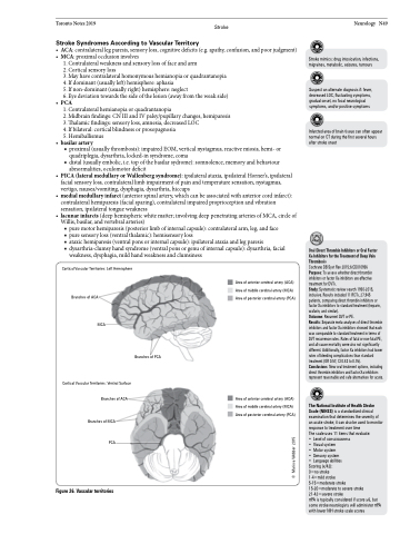
Cortical Vascular Territories: Left Hemisphere
Branches of ACA
MCA
Cortical Vascular Territories: Ventral Surface Branches of ACA
Branches of MCA
PCA
Figure 26. Vascular territories
Area of anterior cerebral artery (ACA) Area of middle cerebral artery (MCA) Area of posterior cerebral artery (PCA)
Branches of PCA
Area of anterior cerebral artery (ACA) Area of middle cerebral artery (MCA) Area of posterior cerebral artery (PCA)
© Marissa Webber 2015
